Bone Cross Section Slide Labeled / File:Ascaris Stained Microscope Slide Labeled Reproductive ... - A flat bone is characterized by parallel surfaces of figure figure observe a slide preparation labeled ground bone;
Bone Cross Section Slide Labeled / File:Ascaris Stained Microscope Slide Labeled Reproductive ... - A flat bone is characterized by parallel surfaces of figure figure observe a slide preparation labeled ground bone;. Some, mostly older, compact bone is remodelled to form these haversian systems (or osteons).the osteocytes sit in their lacunae in concentric rings around a central haversian canal (which runs longitudinally).the osteocytes are arranged in concentric rings of bone matrix called lamellae (little plates), and their processes run in interconnecting canaliculi. In the center of each osteon is the central canal, a space that houses blood vessels and nerves that supply bone. At this level of magnification, the fundamental structure of compact bone is visible. This slide contained a cross section of a very small bone, and you are looking at the entire thickness of the shaft of the bone. Bone cross section slide labeled.
12 photos of the bone cross section labeled. Bone cross section slide labeled. Browse 4,275 bone cross section stock photos and images available, or search for human bone cross section to find more great stock photos and pictures. The section has been ground and dried, hence the lacunae… bone cross section labeled. Related posts of bone cross section.
Move Your Muscles! - Lesson | Skeletal muscle, Muscle ... from i.pinimg.com 12 photos of the bone cross section labeled. The diaphysis and the epiphysis.the diaphysis is the tubular shaft that runs between the proximal and distal ends of the bone. Cross section of a bone labeled. Bone cross section slide labeled. This slide contained a cross section of a very small bone, and you are looking at the entire thickness of the shaft of the bone. Bone is found in the shafts of long bone and consists of various cylindrical units named as haversian system 47. That's why the color looks different. Bone tissue is one of the main components of the skeletal system (other components include bone marrow/marrow cavity, collagen fibers etc).
This section will examine the gross anatomy of bone first and then move on to its histology.
Bone · february 15, 2021. See labeled cross sections of the human body now at kenhub. Mammal compact bone slide, ground c.s. Bone cross section slide labeled. Bone tissue is one of the main components of the skeletal system (other components include bone marrow/marrow cavity, collagen fibers etc). The structure of a long bone allows for the best visualization of all of the parts of a bone ().a long bone has two parts: This slide contained a cross section of a very small bone, and you are looking at the entire thickness of the shaft of the bone. At this level of magnification, the fundamental structure of compact bone is visible. Before placing your slide on the microscope stage, remember to read the label, examine the slide with your eye and note any visible macroscopic features that might help your examination. In this image the bar indicates the location of decalcified compact bone. We obtained 24 axial slices of the normal brain. Learn vocabulary, terms and more with flashcards, games and other study tools. It can be found under the periosteum and in the diaphyses of long bones, where it provides support and protection.
Medical mechanism of action animation reel. Draw and label a cross section of a bone. Bone cross section diagram stretched canvas print | zazzle. See help for more information. Bone cross section slide labeled.
Decalcified Bone Cross Section Labeled / Pathology Of Bone ... from www.researchgate.net Labeled compact bone microscope slides | labeled histology slides. Virtual slide list for histology course. Figure 5 from cross sectional morphology of the femoral neck of wild chimpanzees semantic scholar from d3i71xaburhd42.cloudfront.net. We obtained 24 axial slices of the normal brain. This is a cross section through. 12 photos of the bone cross section labeled. To prepare this slide, a bone specimen is ground thin and then mounted to the slide. Learn vocabulary, terms and more with flashcards, games and other study tools.
See help for more information.
General structure of a mammalian bone. Bone cross section slide labeled. Bone · february 15, 2021. Like other tissues in the body, bones are made up of specialized cells that serve different functions. Note the location of the bone. Mammal compact bone slide, ground c.s. Cross section of bone labeled / exercise 8 cross section of ground compact bone flashcards quizlet / solved chapter 6 quz label the photomicrograph of compect. The compact bone is made up of osteon. From wikimedia commons, the free media repository. 12 photos of the bone cross section labeled. Elongated cylinder orient runs through the core of each osteon. A hand drawn sketch by dr. We obtained 24 axial slices of the normal brain.
Bone tissue and cells under the microscope introduction. Bone is found in the shafts of long bone and consists of various cylindrical units named as haversian system 47. We can see there are two layers of compact bone here. At this level of magnification, the fundamental structure of compact bone is visible. Bones of ankle and foot on x ray 12 photos of the bones of ankle and foot on x ray , bone.
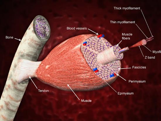
'Cross Section of a Skeletal Muscle Attached to a Bone ... from imgc.allpostersimages.com Copmressive strength for bone is 170×106n/m2. Notice the layered effect in the matrix. Same bone section (slide 7) at higher magnification. Draw and label a cross section of a bone. It can be found under the periosteum and in the diaphyses of long bones, where it provides support and protection. Mammal compact bone slide, ground c.s. Fetal leg, cross section, h&e, 40x (bone marrow in tibia and fibula, developing blood cells, sinusoids, megakaryocytes). Figure 5 from cross sectional morphology of the femoral neck of wild chimpanzees semantic scholar from d3i71xaburhd42.cloudfront.net.
Fetal leg, cross section, h&e, 40x (bone marrow in tibia and fibula, developing blood cells, sinusoid 37829 x 41067, megakaryocyte 37861 x 39647, 38143 x 39087, 39555 x 36969, 31707 x 18214).


0 Komentar