Anatomy Of Ribs Posterior - (2) Thorax I Flashcards | Quizlet - Medial interchondral ligament of right seventh and eighth ribs.. Represents the anatomy of the ribs and muscle attachments. The ribs form the main structure of the thoracic cage protecting the thoracic organs, however their main function is to aid respiration3. However, they do not attach directly to the sternum anteriorly, and instead, attach to the. It branches from the ileocolic artery and may branch further to the appendicular artery. Roughly speaking, this is the area of the chest.
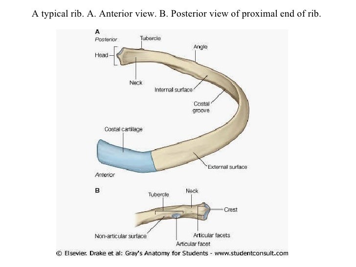
These videos are for educational purpose only for the medical students like. Each rib articulates posteriorly with two thoracic vertebrae by the costovertebral joint. They are twelve in number on either side; The lumbar plexus and its branches. by henry vandyke carter, henry gray (1918) anatomy of the human body. Head, neck, tubercle, and body of a rib.
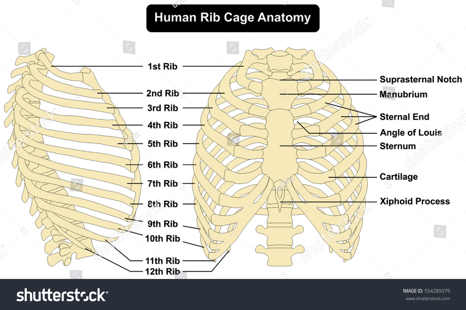
Human Rib Cage Anatomy Anterior Right Stock Vector ... from image.shutterstock.com The number is the same in both males and females. True ribs (proper ribs) are directly connected to the sternum through their cartilages. Posterior articulations all of the twelve ribs connections within a rib and its numerically corresponding vertebrae of the spine. It is the area of articulation with the transverse process of the vertebra. These videos are for educational purpose only for the medical students like. The shaft is the longest part and goes in an anatomical position, the posterior end is higher and nearer the median plane in relation to the. 1.3 ribs anatomy and somatic dysfunctions. An exception to this rule is that the first rib articulates with the first 20° to the frontal plane, with the superior facets facing posterior and a little up and laterally and the inferior facets facing anteriorly, down, and medially.
They are twelve in number on either side;
All 12 pairs of ribs attach to the building blocks of the spine (vertebrae) in the back. Further details of its anatomical relations and muscle attachments can be found in its own section in this text. The posterior end is composed of head, neck, and tubercle. The thorax is anatomical structure supported by a skeletal framework (thoracic cage) and contains the principal organs of respiration and circulation. Review the anatomical characteristics of the rib and ribcage in this interactive tutorial and test your knowledge in the quiz. Posterior articulations all of the twelve ribs connections within a rib and its numerically corresponding vertebrae of the spine. Represents the anatomy of the ribs and muscle attachments. Posteriorly, the heads of the ribs interdigitate with the vertebrae and are numbered according to the inferior vertebra. In this video, you will learn the bony features of typical and atypical ribs. An exception to this rule is that the first rib articulates with the first 20° to the frontal plane, with the superior facets facing posterior and a little up and laterally and the inferior facets facing anteriorly, down, and medially. The lumbar plexus and its branches. by henry vandyke carter, henry gray (1918) anatomy of the human body. Each rib articulates posteriorly with two thoracic vertebrae by the costovertebral joint. Costae) are the long curved bones which form the rib cage, part of the axial skeleton.
Skeletal system anatomy and physiology nurseslabs. Posterior left rib fractures with injuries and nonunion of. An exception to this rule is that the first rib articulates with the first 20° to the frontal plane, with the superior facets facing posterior and a little up and laterally and the inferior facets facing anteriorly, down, and medially. Common characteristics of the ribs figs. Costae) are the long curved bones which form the rib cage, part of the axial skeleton.
Lecture 1 thoracic wall from image.slidesharecdn.com Common characteristics of the ribs figs. This incision may be continued across the costal margin to open the abdominal cavity as in. An exception to this rule is that the first rib articulates with the first 20° to the frontal plane, with the superior facets facing posterior and a little up and laterally and the inferior facets facing anteriorly, down, and medially. Posterior left rib fractures with injuries and nonunion of. Both muscles attach to various ribs and parts of the spine. Illustrations in anterior and posterior view of male torso and back, allowing the lines and regions used in surface anatomy to be displayed (midclavicular line, midline, pectoral region, sternal region.) ribs: In the anatomical position, the scapula overlies the second to seventh ribs on the posterolateral aspect of the chest wall. Be sure to subscribe to the visible body blog for more anatomy awesomeness!
Each pair articulates with a different thoracic vertebra on the posterior side of the body.
The nomenclature of the costal veins is the same as the arteries. Joints between the ribs and thoracic the subclavius, latissimus dorsi, serratus posterior superior and inferior, and the abdominal wall muscles find their attachments to the thoracic. The most superior rib is designated rib 1 and it articulates with the t1 thoracic vertebrae. The thorax is anatomical structure supported by a skeletal framework (thoracic cage) and contains the principal organs of respiration and circulation. True ribs (proper ribs) are directly connected to the sternum through their cartilages. Each pair articulates with a different thoracic vertebra on the posterior side of the body. Head, neck, tubercle, and body of a rib. Includes images, video, and free quiz. Posteriorly, the heads of the ribs interdigitate with the vertebrae and are numbered according to the inferior vertebra. Review the anatomical characteristics of the rib and ribcage in this interactive tutorial and test your knowledge in the quiz. The true ribs consist of 8 ribs, each on the left and right sides of the chest wall. Further details of its anatomical relations and muscle attachments can be found in its own section in this text. The ribs are elastic arches of bone, which form a large part of the thoracic skeleton.
Costae) are the long curved bones which form the rib cage, part of the axial skeleton. Be sure to subscribe to the visible body blog for more anatomy awesomeness! The ribs are elastic arches of bone, which form a large part of the thoracic skeleton. Both muscles attach to various ribs and parts of the spine. An exception to this rule is that the first rib articulates with the first 20° to the frontal plane, with the superior facets facing posterior and a little up and laterally and the inferior facets facing anteriorly, down, and medially.
Introduction & Anatomy Thoracic — The Gap Physio from static1.squarespace.com They are twelve in number on either side; Skeletal system anatomy and physiology nurseslabs. The ribs form the main structure of the thoracic cage protecting the thoracic organs, however their main function is to aid respiration3. The ribs, along with the thoracic vertebrae, sternum, and costal cartilages. Each rib articulates posteriorly with two thoracic vertebrae by the costovertebral joint. They articulate with the vertebral column posteriorly, and terminate anteriorly as cartilage (known as costal cartilage). The posterior abdominal wall is a musculoskeletal structure formed by the posterior abdominal muscles, their fascia, the lumbar vertebrae and the image: The thorax is anatomical structure supported by a skeletal framework (thoracic cage) and contains the principal organs of respiration and circulation.
Includes images, video, and free quiz. The subclavian artery and brachial plexus cross the rib posterior to anterior scalene muscle attachment and then run in contact with the bone on their way to the upper limb. Anatomical name for floating ribs, anatomical term ribs, anatomical word for ribs, anatomy ribs quiz, ribs anatomical position, human anatomy anatomy of shoulder 12 photos of the anatomy of shoulder anatomy of nerves in shoulder, anatomy of posterior shoulder dislocation, anatomy. They articulate with the vertebral column posteriorly, and terminate anteriorly as cartilage (known as costal cartilage). It branches from the ileocolic artery and may branch further to the appendicular artery. Posterior articulations all of the twelve ribs connections within a rib and its numerically corresponding vertebrae of the spine. Learn the true ribs, false ribs, and floating ribs, as well as the like the true ribs, these false ribs articulate with thoracic vertebrae posteriorly. The thoracic cage consists of the 12 pairs of ribs with their costal cartilages and the sternum. Posteriorly, the heads of the ribs interdigitate with the vertebrae and are numbered according to the inferior vertebra. The costotransverse ligaments in human: Made up of thoracic vertebrae, ribs and… functions at upper end to connect the shoulder girdle and conn… Major landmarks of a typical rib are the following: Medial interchondral ligament of right seventh and eighth ribs.
The most superior rib is designated rib 1 and it articulates with the t1 thoracic vertebrae anatomy of ribs. Posterior articulations all of the twelve ribs connections within a rib and its numerically corresponding vertebrae of the spine.


0 Komentar